Analysis methods / Research
My examination of stringed instruments is exclusively non-invasive. I work as gently as possible with the instrument at all times, for the most part even without contact. My primary tool for these examinations is a high-resolution digital camera, which, due to its setting options, goes beyond human vision and generates visualisations.
The following comparative images were created with the same violin (David Tecchler, Rome, 1722) in different light spectra.
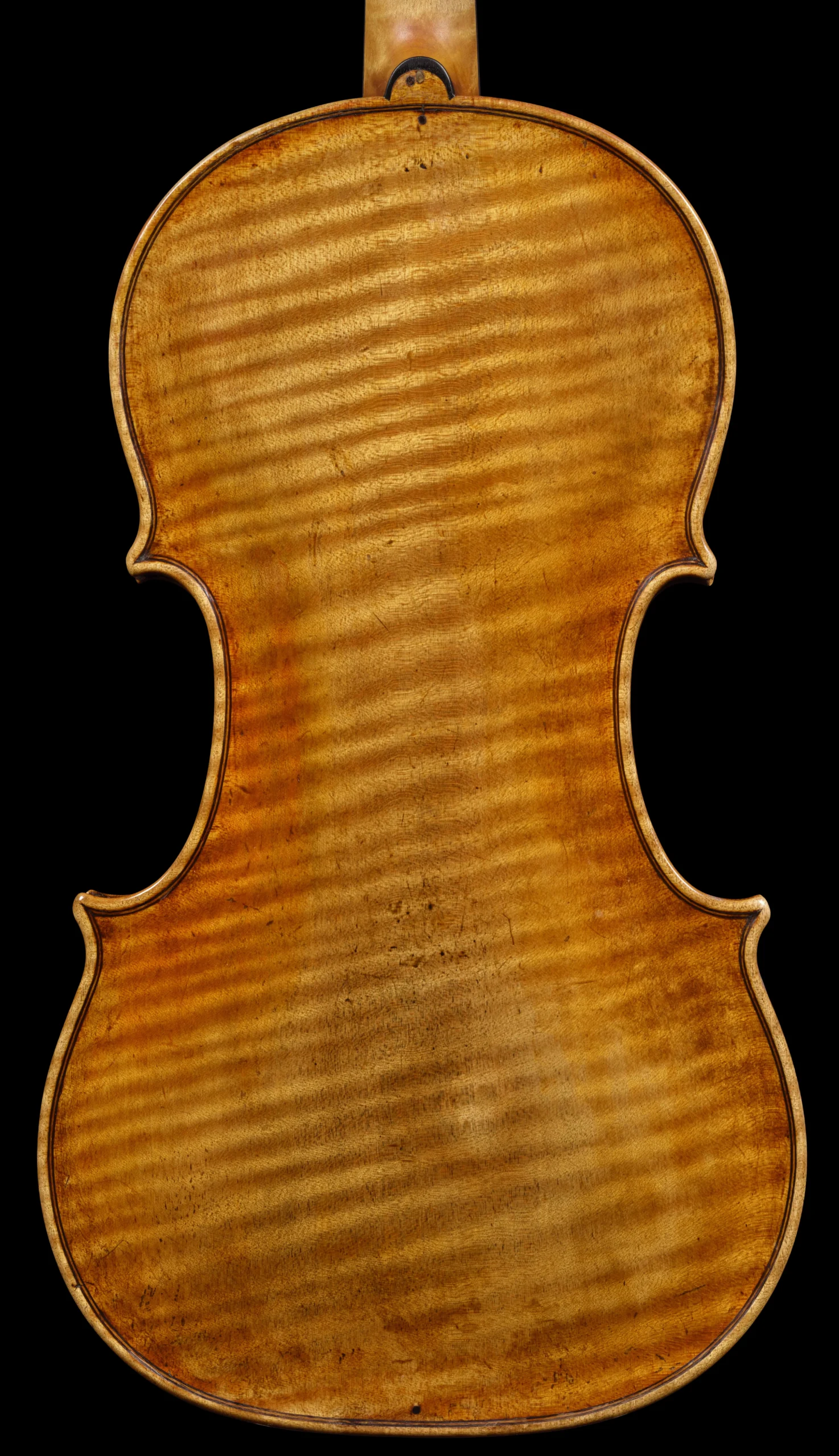

UV light (320- 400nm)
left: daylight (VIS); right: UV light.
The most common light analysis method used in this field to examine the varnish and surface of stringed instruments is in the ultraviolet range between 320- 400nm.The image of the resulting fluorescence enables the trained observer to distinguish retouching and other additions from the original. This examination forms the basis of an extensive analysis with light.
Cyan light (490 nm)
left: UV light; right: cyan light
In the article of the publication “Antonio Stradivari ‘Barjansky’ Cello” (Jost Thöne Verlag 2021) I presented, together with Prof. Dr. Martin Bek, another important wavelength for the examination of stringed instruments. It can provide additional, important information about the condition of the varnish. The light is at a wavelength of 490 nm (cyan light) and, in simple terms, causes the original (coloured) varnish of the instrument to fluoresce. The properties of the resulting fluorescence are considerably more transparent, i.e. less opaque, than in UV light. It is therefore possible to identify original varnish even in the presence of additions and retouching. This can be an important component for a planned restoration.
As the study of this wavelength is still very new, it is not yet possible to define exactly which substance or mixture of substances elicits these fluorescences. However, there are indications that it could be the oils in the varnish.


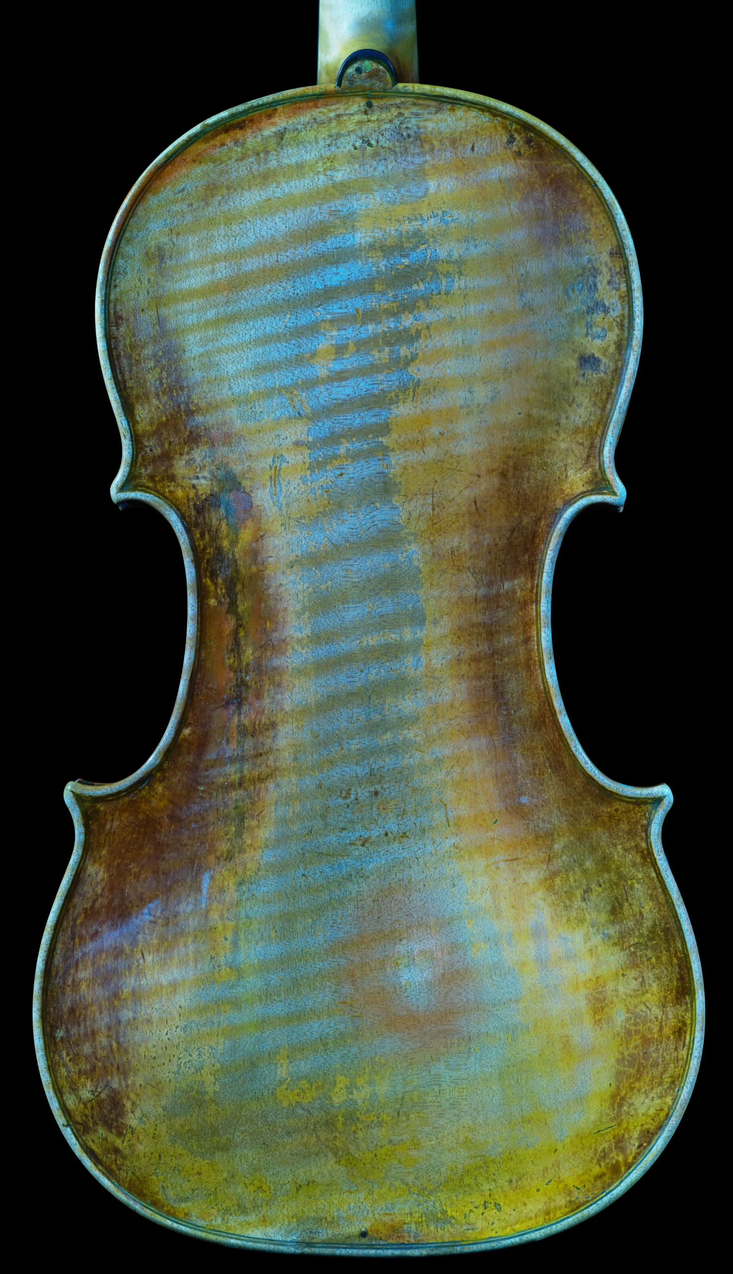
Blue light (460 nm) with optical filter
left: cyan light; right: blue light
Since some relevant fluorescences, such as those of coating varnishes, are less pronounced at the wavelength of 490nm, another, shorter wavelength light at 460nm (blue light) can be just as interesting. For this purpose a special long-pass filter (500nm) is used for the camera. A special feature of this wavelength is that rather unclear areas (those that fluoresce in UV or cyan light similarly to the original varnish, or in an indefinably different way) can be better defined by way of comparison.
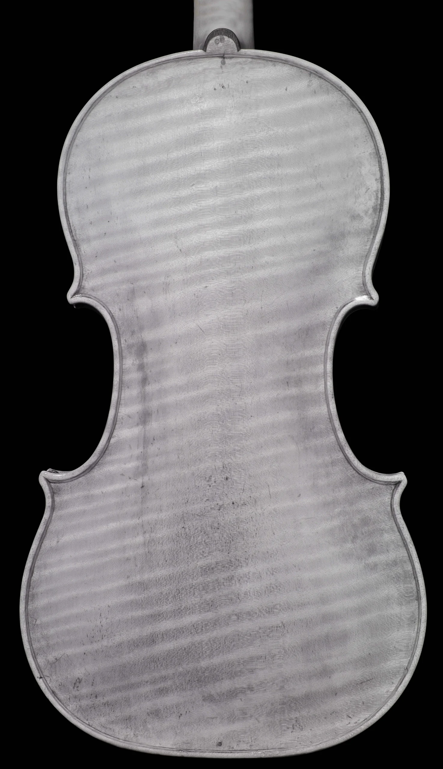
Infrared light (incident light, 940nm)
left: daylight; right: infrared incident light
Examination with infrared light, which is invisible to the human eye, is particularly valuable when examining paintings. Underlying layers can be made visible with the right equipment. Of course, this is very helpful in understanding how the painting was created and reveals many hidden details.
This can be just as interesting with stringed instruments. Especially retouching, patina, and some additions can be seen more clearly. However, a view into deeper layers is not achieved, or not to the extent hoped for.
Analyses in infrared light and other spectral wavebands are normally projected onto the object in incident light during art analysis. However, as you will see in the next examination method, this can also be done differently with this light.


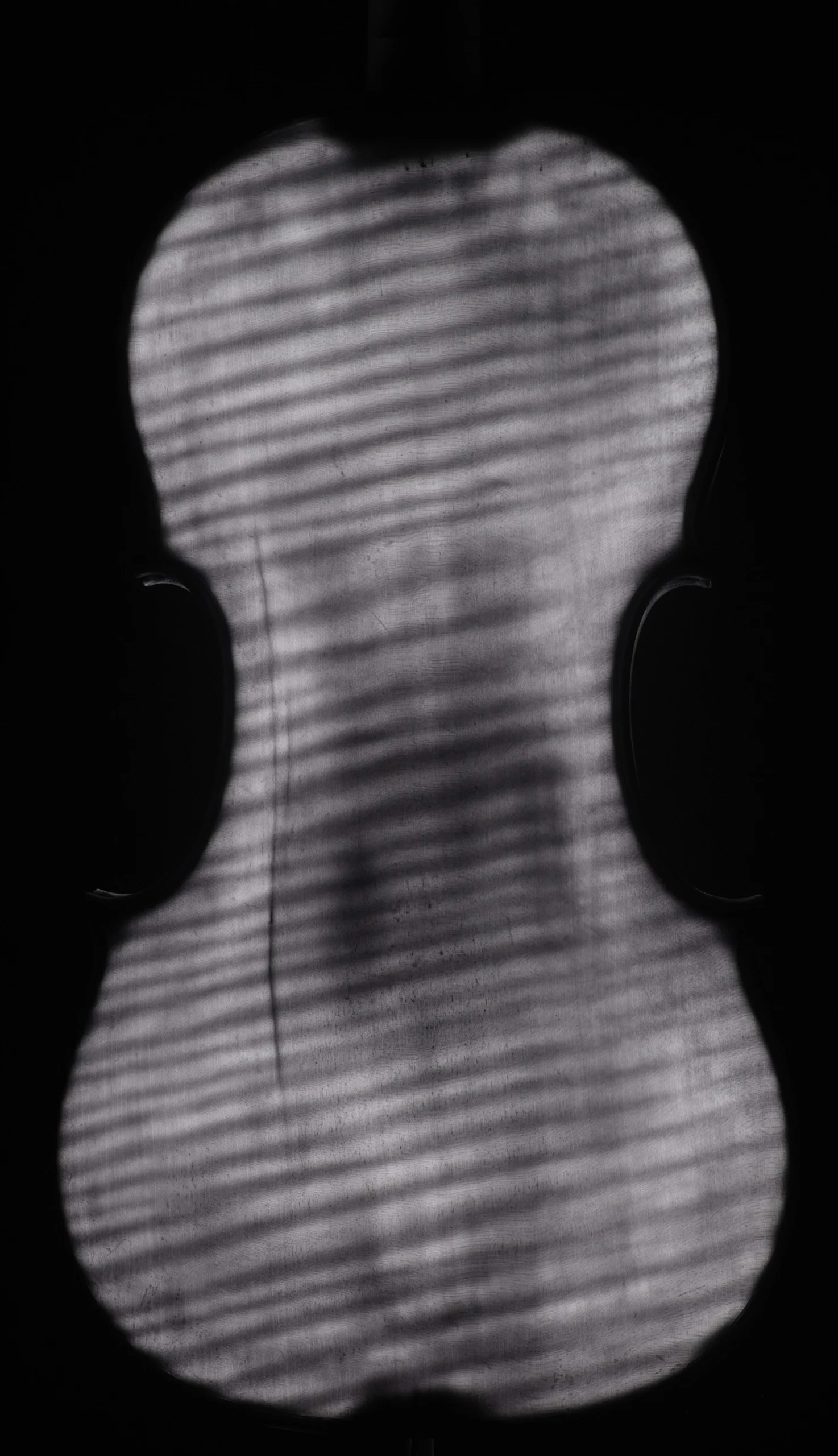
IR scan (infrared transmitted light, 940nm)
left: daylight; right: Infrared transmitted light
Because of the strong permeability of the wood to infrared light, a different (than in the previously described IR examination method), rather unconventional set-up of the light source and camera is also possible and makes sense. I place the light source inside the body and take pictures from the outside with a multispectral, infrared-capable camera, so that an image is produced that most closely resembles an X-ray.
Damage such as cracks, fractures, worm damage, internally glued inlays, lining, the position of the bass bar and soundpost as well as growth forms of the wood are fantastically visible with this “infrared scan“.
This method has been published by me in the professional journal TheStrad (June 2022).
If you are in the process of buying and want to make sure that the condition of the wood meets your expectations, this method is excellent. Due to the relatively low costs compared to an equally useful CT scan,
such an examination is already worthwhile with an instrument valued at about 10,000 euros.
Dendrochronological examination
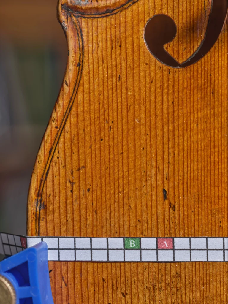
Through cooperation with internationally recognised dendrochronologists, it is also possible in my workshop to carry out a dendrochronological examination of the top wood (spruce). I will take the necessary high-resolution pictures of the top of your instrument and forward them to the specialists (e.g. Arjan Versteeg). The result will then either be communicated verbally or, if desired, an expertise will be drawn up.
This method of examination is particularly attractive because it is one of the very few objective scientific examinations in stringed instrument analysis. You usually receive the result of the age of the top wood as well as correlations with other measurements from the extensive database of colleagues.
Just like the IR scan, this examination is highly recommended when buying or selling an instrument in order to avoid surprises.
Documentation of arching processes
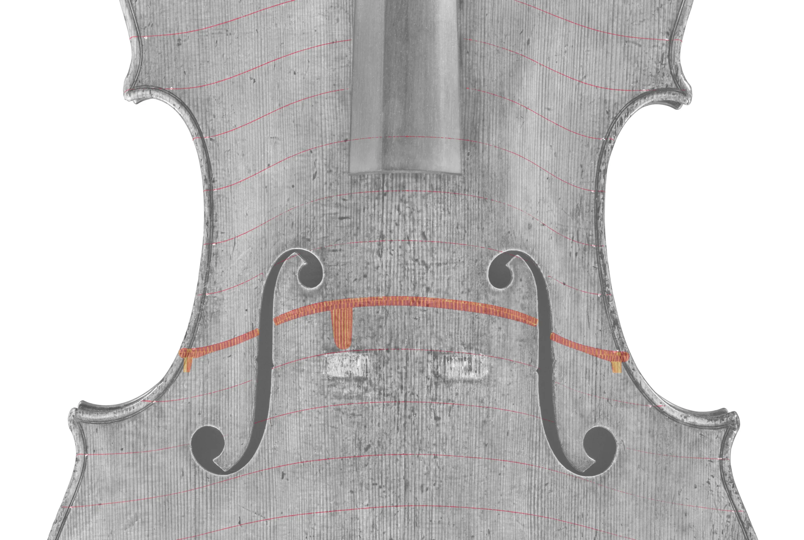
Measuring the contours of the arching of the top and back of historically valuable stringed instruments is an important part of the documentation process and can, for example, provide evidence of changes to the instrument.
The method used for this purpose, which I presented for the first time in the monograph “Antonio Stradivari ‘Barjansky’ Cello”, is based on a laser line projected at right angles onto the arching to be measured and a digital camera mounted on a bellows device. The image plane of the camera is positioned parallel to the projection direction of the laser line and aligned diagonally from above in such a way that it is also possible to view areas of the middle bout.
Despite the oblique angle of view of the instrument, the curvature indicated by the laser is displayed without distortion with this technique.
